Happy Clients

Card image cap

Schatzkar’s type 5 fractures

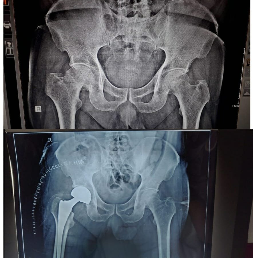
The patient is a 28-year-old health-conscious bank employee who enjoys playing football during weekends. During one such weekend, he was unfortunately tackled in an aggressive manner which landed a kick directly to his left knee. 

The patient recalls falling down with intense pain in his knee joint; his knee started swelling very soon after the injury. In the following days, the pain only worsened, and he was unable to put any weight on the injured leg. He was then referred to our Wholistic Care Centre for consultation with Dr. Saurabh Talekar.


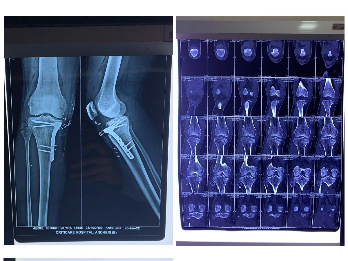
Proximal tibia fractures are common, and Schatzkar’s type 5 fractures are challenging to fix. Hence performing a CT scan is a standard protocol for planning the surgery, and MRI scans are typically not performed.  However, we performed the MRI scan due to high clinical suspicion, and a medial meniscal tear was found, which was treated in the same sitting through Arthroscopy. The ORIF and meniscal repair was hence done in the same setting.


Before the surgery, the highly-skilled Dr. Saurabh Talekar discussed the risks, benefits, and potential complications with the patient and his family. The patient was given spinal anaesthesia during the surgery which avoids complications of General Anaesthesia and at the same time provides Adequate anaesthesia. During the operation, Dr. Saurabh Talekar and his team carefully monitored the patient’s vital signs, like his blood pressure and heart rate.


Dr. Talekar first cleaned the injured area and made an incision through the skin and muscle. Then he brought the pieces of the patients’ tibia back into alignment. Next, he secured the pieces of the tibia to each other (known as fixation). Fixation is a process where the surgeon uses tools like nails, screws, metal plates, wires, or pins. The Meniscus was repaired through a minimally invasive approach without entering the joint. The procedure was performed by visualisation through an arthroscope into the joint.

Card image cap

Hemiarthroplasty

Patient Kishore Moon consulted Dr. Saurabh Talekar, an orthopedic surgeon in Kandivali, Mumbai, at Wholistic Care Centre with severe hip pain. He had a fall at home, due to which he was unable to walk and was bedridden.

Initially, the doctor performed a physical examination, discussed the medical history, and asked the patient to undergo an x-ray. The x-ray showed a fracture of the femur neck. After analysing the x-ray and the patient’s overall health, Dr. Saurabh Talekar suggested hemiarthroplasty. 

He explained the benefits of arthroplasty to the patient and his family, and they gave their consent for the procedure. 

Hemiarthroplasty(partial hip replacement)

Hip hemiarthroplasty is a procedure that replaces a part of the hip. It is generally performed in cases of traumatic accidents where the femoral head is fractured, like high-impact falls that dislocate and fracture the hip.

Hemiarthroplasty requires less surgical time, and there is less loss of blood. Further, the risk of dislocating the hip after the surgery is also minimum. Neck of femur fractures make patients bedridden. 

Even at an advanced age it is recommended to operate and mobilise the patient out of bed as early as possible to avoid complications related to long term bed rest. 

Complications of non-operative treatment can be Urinary track infections, Lung infections, bedsores, deep vein thrombosis, pulmonary embolism.

Surgical procedure

After completing all the pre-operative tests, the patient Kishore Moon was cleared for the procedure. He was administered an epidural that made his lower region numb while awake.

During the procedure, Dr. Saurabh Talekar, an experienced orthopedic surgeon in Kandivali, Mumbai, replaced the fractured femur head with an implant. He detached the femoral head from the femur and hollowed out the inside of the femur. Then he inserted a metal stem firmly inside the femur. Finally, he closed the incision with sutures and bandaged it.

Recovery

The surgery was completed without any complications, and the doctor prescribed pain medications and other medicines for quick recovery.

On the second day after the surgery, the patient was made to walk. He was started with physical therapy in the hospital, which he had to continue even after discharge. Physical therapy is necessary for regaining flexibility and strength in the hip. 

Patient Kishore Moon had to visit Wholistic Care Centre for follow-ups so that Dr.Saurabh Talekar could monitor his progress.

Card image cap

Fracture Proximal Humerus

A female patient with severe shoulder and arm pain and deformity visited Dr.Saurabh Talekar, an orthopedic surgeon in Kandivali, Mumbai, at Wholistic Care Centre.

She had a fall due to which there was localized pain in the arms and shoulder and a limited range of motion. She was unable to raise her arm or perform any activity.

She had consulted another doctor who had recommended surgical intervention.


Assessment

Dr. Saurabh Talekar did a physical examination, and X-rays were taken. X-ray is the standard diagnostic method to verify a fracture, and mainly two different angles of view are taken to help analyse the pattern of the fracture. The x-ray showed a fracture in the proximal humerus.

After clinical assessment, Dr. Saurabh Talekar suggested a conservative treatment using a hanging cast to address the fracture.


Conservative treatment for fractures

Conservative fracture treatment is not quite common nowadays, with orthopedic surgeons and patients opting for surgical interventions. 

However, conservative fracture treatment offers less trauma and complications. and the results are similar to the operative method.

Further, the cost of the treatment is considerably less as it does not require surgery. 

Traditional conservative fracture management is based on three main principles: fracture reduction, fracture stabilisation, and keeping the fracture stabilised with a cast or splint until it heals.

Using conservative treatment with a hanging arm cast requires accuracy and precise attention to detail. The orthopedic surgeon will reposition the fragmented bone, close the wound if necessary and then apply a cast or splint to secure the bones in place.

Immobilisation helps to reattach the bones and aid with the healing process. The doctor tries for early mobilisation to avoid joint stiffness.


Treatment provided

With the patient’s consent, Dr. Saurabh Talekar, a prominent orthopedic surgeon in Kandivali, Mumbai, went ahead with the conservative treatment of the fracture. He placed the arm in a hanging cast.

He called the patient for regular follow-ups to monitor the progress. After a week, he asked for an x-ray, which showed signs of healing.

We took an x ray to confirm reduction at one week. At 3 weeks there were signs of consolidation of the fracture

Then the cast was removed, and the patient continued using slings for another three weeks. Dr. Saurabh Talekar asked her to move the hand in a pendulum movement.

During the seventh week, the patient could move the arm behind her back and perform internal rotation during the follow-up treatment.

Book an Appointment

Enter details,our team would approach to help you as soon as possible.

Phone icon
Call Now